Monocyte differentiation and polarization are widely used to generate macrophage and dendritic cell (DC) models for studying innate immune responses. However, experimental workflows can vary considerably depending on the cell model and differentiation or polarization conditions. In this application note, we demonstrate how InvivoGen’s M1 and M2 Polarization Mixes and DC Differentiation Mix provide standardized workflows adapted to human peripheral blood CD14+ monocytes and THP-1 cells.
From monocytes to macrophage and DC models
Monocytes are innate, circulating immune cells that can differentiate into macrophages or DCs in response to specific environmental signals and local tissue conditions. In vitro, monocytes can differentiate into M0 macrophages and subsequently acquire different functional states, including M1- and M2-like phenotypes1,2. M1 macrophages are associated with pro-inflammatory and antimicrobial responses, whereas M2 macrophages are associated with immunoregulation, inflammation resolution, and tissue repair1,3. In parallel, DCs are specialized antigen-presenting cells that capture and process antigens, present antigen-derived peptides to T cells, and contribute to the regulation of adaptive immune responses4,5. Altered macrophage polarization and DC function have been associated with a broad range of inflammatory and autoimmune diseases (e.g., rheumatoid arthritis and systemic lupus erythematosus), as well as cancer1,6,7.
Two complementary human monocytic models are commonly used to investigate these processes and better understand the mechanisms underlying immune cell differentiation and function. Human peripheral blood CD14⁺ monocytes provide a physiologically relevant primary model but can be limited by donor-to-donor variability and restricted cell availability, which depends on access to human blood samples and the number of monocytes that can be isolated2,4. In contrast, THP-1 monocytes are immortalized cells derived from a leukemia patient and therefore do not fully mimic the physiological characteristics of primary monocytes. Nevertheless, they provide a renewable and more homogeneous human cell model, facilitating standardized experiments, genetic manipulation, and reporter-based functional studies. However, differentiation and polarization protocols can vary considerably between studies, particularly when using THP-1 cells2,5,8,9.
Standardized differentiation and polarization
To facilitate differentiation and polarization across both models, InvivoGen has developed M1 Polarization Mix, M2 Polarization Mix, and a DC Differentiation Mix, with dedicated protocols for human peripheral blood CD14⁺ monocytes and THP-1 cell lines (Figure 1).
Based on established differentiation and polarization signals, these formulations combine defined cytokines and other factors to reduce experimental variability and improve reproducibility. Each mix undergoes lot-to-lot validation to ensure consistent performance across experiments. The underlying mechanisms of macrophage polarization and DC differentiation are described below:
Macrophage polarization
For macrophage studies, human peripheral blood CD14⁺ monocytes are first differentiated into M0 macrophages using M-CSF, whereas THP-1 cells are differentiated using PMA2,8,9. The resulting M0 macrophages provide a common starting point for subsequent M1 or M2 polarization.
M1 Polarization Mix combines IFN-γ and LPS, two complementary inflammatory signals commonly used to induce M1-like macrophage polarization1–3. IFN-γ activates JAK/STAT1 signaling, while LPS engages TLR4 and activates inflammatory signaling pathways, including NF-κB and MAPKs. Together, these signals promote transcriptional programs associated with inflammatory and antimicrobial macrophage functions and the acquisition of an M1-like phenotype.
M2 Polarization Mix combines IL-4 and IL-13, two cytokines commonly used to induce alternative macrophage polarization. Both cytokines signal through IL-4Rα receptor complexes and converge on JAK/STAT6 signaling, promoting transcriptional programs associated with immunoregulatory and tissue-repair functions characteristic of M2-like macrophages1–3.
DC differentiation
DC Differentiation Mix combines GM-CSF, IL-4, TNF-α, and ionomycin. GM-CSF and IL-4 are commonly used to drive monocytes toward a DC phenotype through signaling pathways, including JAK/STAT5 and JAK/STAT6, respectively, while additional maturation signals promote the acquisition of DC-associated properties4,5. TNF-α provides a pro-inflammatory maturation signal, whereas ionomycin increases intracellular Ca²⁺ levels, activating calcium-dependent signaling pathways that contribute to DC activation and maturation. Notably, the combination of GM-CSF, IL-4, TNF-α, and ionomycin has been described for the generation of mature DC-like cells from THP-1 cells5.
Characterization of M1 and M2 macrophages and DCs
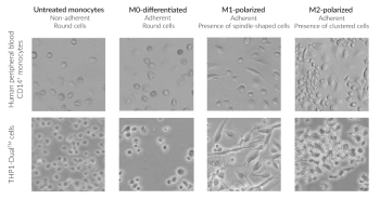
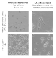
Monocyte differentiation and polarization are accompanied by changes in cell morphology that reflect the acquisition of different cellular phenotypes2,3,8,9. As shown in Figure 1, THP-1 cells display distinct morphological changes following M0 differentiation and subsequent M1 or M2 polarization, as well as following DC differentiation. Similar morphological changes were observed with human peripheral blood CD14⁺ monocytes (data not shown).
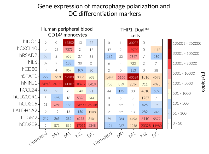
Gene expression analysis provides complementary molecular characterization of the generated populations. Digital PCR (dPCR) was used to assess the expression of markers associated with M1 macrophages (e.g., CXCL10, IDO1), M2 macrophages (e.g., CD200R1, TGM2), and DCs (e.g., CD209), represented as a heatmap (Figure 2). Rather than relying on morphology alone, combining morphological observations with gene expression and/or phenotypic analyses provides complementary evidence of the differentiation and polarization states obtained with each protocol1–5,8,9.
Together, InvivoGen’s M1 and M2 Polarization Mixes and DC Differentiation Mix provide standardized workflows for generating distinct macrophage and DC populations from both primary and immortalized human monocytes. The use of dedicated protocols for each cell model facilitates their parallel characterization and comparison, providing a common experimental framework for studying monocyte differentiation and polarization.
InvivoGen also provides reporter THP-1 cell lines for the functional assessment of immune signaling following differentiation and polarization. For example, THP1-Dual™ cells feature dual NF-κB and ISG reporter systems, allowing pathway activation to be monitored in polarized macrophages or differentiated DCs. Knockout (KO) and knockin (KI) THP-1 cell lines can further be used to investigate the contribution of specific genes to these responses.
Lear more information about THP-1 cell lines
References:
1. Luo M. et al., 2024. Macrophage polarization: an important role in inflammatory diseases. Front. Immunol. 15:1352946.
2. Mohd Y.Z. et al., 2022. Macrophage polarization in THP-1 cell line and primary monocytes: A systematic review. Differentiation 128:67–82.
3. Orecchioni M. et al., 2019. Macrophage polarization: Different gene signatures in M1(LPS+) vs. classically and M2(LPS−) vs. alternatively activated macrophages. Front. Immunol. 10:1084.
4. Segura E. et al., 2025. Monocyte-Derived Dendritic Cells: An Updated View on an Old Concept. Immunol. Rev.
5. Hölken J.M., Teusch N., 2023. The Monocytic Cell Line THP-1 as a Validated and Robust Surrogate Model for Human Dendritic Cells. Int. J. Mol. Sci. 24:1452.
6. Yamada S. et al., 2025. Dendritic cell heterogeneity and its role in connective tissue diseases. Inflamm. Regen. 45:24.
7. Moon C. et al., 2025. Dendritic cell maturation in cancer. Nat. Rev. Cancer 25(4):225–248.
8. Forrester M.A. et al., 2018. Similarities and differences in surface receptor expression by THP-1 monocytes and differentiated macrophages polarized using seven different conditioning regimens. Cell Immunol. 332:58–76.
9. Klatt S. et al., 2026. Comparison of THP-1 Subtypes Reveals Differences on Transcriptomic and Protein Expression Level Throughout Their Immunological Differentiation. J. Immunol. Res. 2026:e9427482.