


DC Differentiation Mix
-
Cat.code:
mix-dcNEW
- Documents
ABOUT
Mix for reproducible dendritic cell differentiation of human monocytes
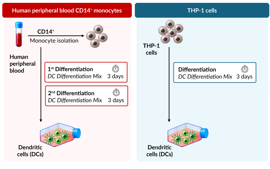
DC Differentiation Mix is a ready-to-use formulation supplied with a standardized protocol for reproducible dendritic cell (DC) differentiation of primary human monocytes or the human THP-1 monocytic cell line:
- Primary human CD14⁺ monocytes: differentiate into DCs with two consecutive treatments of DC Differentiation Mix.
- Human THP-1 cells: differentiate into DCs with DC Differentiation Mix.
The mix combines GM-CSF, IL-4, TNF-α, and ionomycin, which drive DC differentiation through complementary pathways: GM-CSF activates JAK2/STAT5, PI3K/AKT, and MAPK signaling, IL-4 activates JAK/STAT6 signaling, TNF-α activates NF-κB and MAPK signaling, and ionomycin increases intracellular Ca²⁺ signaling. After treatment, differentiated cells from both sources display a characteristic DC morphology, with the presence of round non-adherent cells and branched adherent cells, and express human DC-associated markers, including cell-surface CD209 (DC-SIGN) and intracellular ALDH1A2 (see figures).
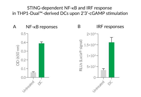
THP-1 cells provide a convenient model for studying DC differentiation and function. Reporter cell lines such as THP1-Dual™ allow functional assessment of DC-differentiated cells via two readouts: NF-κB-inducible SEAP and ISG-inducible Lucia® luciferase (see figures).
Applications
- Generation of DC models from human monocytes
- Functional characterization of DC-differentiated cells
- Evaluation of immunomodulatory compounds targeting macrophage polarization and function
Dendritic cells are antigen-presenting cells that play a central role in linking innate and adaptive immunity through antigen presentation and T-cell activation. Their study can rely on complementary models: primary monocytes provide greater physiological relevance, while immortalized monocytic cells offer a renewable and standardized model that facilitates genetic manipulation and KO/KI studies.

All InvivoGen products are for internal research use only, and not for human or veterinary use.
InvivoGen also offers:
SPECIFICATIONS
Specifications
See Technical Data Sheet (TDS)
The absence of bacterial contamination (e.g. lipoproteins and endotoxins) has been confirmed using HEK-Blue-Lucia™ hTLR2 and HEK‑Blue-Lucia™ mTLR4 cells
Each lot is validated for successful polarization of THP1-Dual™ cells by morphological assessment using microscopy and cell surface marker analysis by flow cytometry
Each vial of DC Polarization Mix is sufficient for the polarization of 2.5 × 10⁷ cells
CONTENTS
Contents
-
Product:DC Differentiation Mix
-
Cat code:mix-dc
-
Quantity:1 vial
Each vial of DC Polarization Mix is sufficient for the polarization of 2.5 × 10⁷ cells
Shipping & Storage
- Shipping method: Room temperature
- -20°C
- Avoid repeated freeze-thaw cycles
Storage:
Caution:
Details
Monocyte differentiation and polarization
Monocytes are circulating innate immune cells that can differentiate into macrophages or dendritic cells (DCs) in response to environmental signals. Monocytes can differentiate in vitro into M0 macrophages, commonly using M-CSF or GM-CSF, and subsequently acquire different functional states, including M1- and M2-like phenotypes [1,2]. M1 macrophage polarization is promoted by inflammatory signals such as IFN-γ and microbial stimuli such as LPS. M1-like macrophages contribute to antimicrobial and pro-inflammatory responses and produce mediators including TNF-α, IL-1β, IL-6, and IL-12 [1,3]. In contrast, M2 macrophage polarization is promoted by cytokines such as IL-4 and IL-13. M2-like macrophages are associated with immunoregulation, inflammation resolution, and tissue repair and can produce regulatory mediators such as IL-10 and TGF-β [1,3].
DC differentiation in vitro is commonly induced by GM-CSF and IL-4, while additional stimuli can promote their activation and maturation. TNF-α promotes DC maturation and the expression of co-stimulatory molecules, whereas ionomycin, a calcium ionophore, induces intracellular Ca²⁺ signaling and can promote DC activation and maturation [4,5]. The combination of GM-CSF, IL-4, TNF-α, and ionomycin has also been used to generate mature DC-like cells from THP-1 cells [5]. DCs capture and process antigens, present antigen-derived peptides to T cells, and produce cytokines that shape subsequent immune responses [4,5].
Altered macrophage polarization and DC function have been associated with autoimmune and inflammatory diseases, including rheumatoid arthritis and systemic lupus erythematosus, as well as cancer [1,6,7].
Morphological and phenotypic features
Monocyte differentiation and polarization are accompanied by changes in cell morphology and molecular marker expression. M0 macrophages acquire an adherent macrophage-like morphology following differentiation. Further polarization results in distinct morphological and phenotypic features, although these can vary according to the cell source, stimulus, and experimental conditions [2,8,9].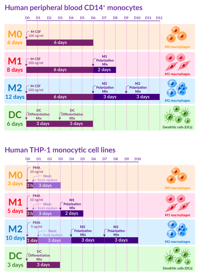
M1-like macrophages can display a spread or spindle-shaped morphology and are commonly associated with markers such as CD80, IDO1, and CXCL10. M2-like macrophages can display elongated or clustered morphologies and are commonly associated with markers CD200R1, CD206, and TGM2 [1–3,8,9]. As macrophage phenotypes are highly plastic, combinations of morphological and molecular markers are generally more informative than individual markers for characterizing polarization states [1,3].
DC differentiation is accompanied by changes in cell adherence and morphology, including the development of cellular projections characteristic of dendritic cells. Their phenotype can be characterized by the expression of DC-associated markers such as CD209 (DC-SIGN), together with changes in other differentiation- and activation-associated markers [4,5].
In vitro models for polarization studies
Human peripheral blood CD14+ monocytes provide a physiologically relevant primary model for studying macrophage polarization and DC differentiation. However, their experimental use can be limited by donor-to-donor variability and cell availability [2,4]. In contrast, THP-1 cells provide a renewable and more homogeneous human monocytic cell model, facilitating standardized experiments and reducing some of the biological variability associated with primary cells. THP-1 cells can be differentiated into macrophage-like cells and subsequently polarized toward M1- or M2-like phenotypes, although differentiation and polarization protocols vary between studies [2,3,8,9]. They can also be differentiated toward DC-like phenotypes, providing a reproducible model for studying DC differentiation and activation [5].
Primary CD14+ monocytes and THP-1 cells therefore provide complementary experimental models: primary cells offer greater physiological relevance, whereas THP-1 cells provide a renewable and standardized system suited to reproducible experiments, genetic manipulation, and reporter-based functional studies [2,5,8,9]. InvivoGen has developed M1 and M2 Polarization Mixes and DC Differentiation Mix, with dedicated protocols adapted to each cell model, enabling standardized generation of M1 and M2 macrophages and differentiated DCs (see figure).
References:
1. Luo M. et al., 2024. Macrophage polarization: an important role in inflammatory diseases. Front. Immunol. 15:1352946.
2. Mohd Y.Z. et al., 2022. Macrophage polarization in THP-1 cell line and primary monocytes: A systematic review. Differentiation 128:67–82.
3. Orecchioni M. et al., 2019. Macrophage polarization: Different gene signatures in M1(LPS+) vs. classically and M2(LPS−) vs. alternatively activated macrophages. Front. Immunol. 10:1084.
4. Segura E. et al., 2025. Monocyte-Derived Dendritic Cells: An Updated View on an Old Concept. Immunol. Rev.
5. Hölken J.M., Teusch N., 2023. The Monocytic Cell Line THP-1 as a Validated and Robust Surrogate Model for Human Dendritic Cells. Int. J. Mol. Sci. 24:1452.
6. Yamada S. et al., 2025. Dendritic cell heterogeneity and its role in connective tissue diseases. Inflamm. Regen. 45:24.
7. Moon C. et al., 2025. Dendritic cell maturation in cancer. Nat. Rev. Cancer 25(4):225–248.
8. Forrester M.A. et al., 2018. Similarities and differences in surface receptor expression by THP-1 monocytes and differentiated macrophages polarized using seven different conditioning regimens. Cell Immunol. 332:58–76.
9. Klatt S. et al., 2026. Comparison of THP-1 Subtypes Reveals Differences on Transcriptomic and Protein Expression Level Throughout Their Immunological Differentiation. J. Immunol. Res. 2026:e9427482.
DOCUMENTS
Documents
Technical Data Sheet
Validation Data Sheet
Safety Data Sheet
Certificate of analysis
Need a CoA ?


 Powered by Bioz