



HEK-Blue™ mIL-4/IL-13 Cells
-
With a free vial of mouse IL-4 protein
-
Cat.code:
hkb-mil413-4NEW
- Documents
ABOUT
Mouse IL-4 & IL-13 responsive STAT6-SEAP reporter assay
HEK-Blue™ mIL-4/IL-13 cells are designed to monitor mouse IL-4 or IL-13-induced STAT6 stimulation or inhibition. This colorimetric bioassay can be used for screening activatory molecules, such as engineered cytokines, or inhibitory molecules, such as neutralizing antibodies.
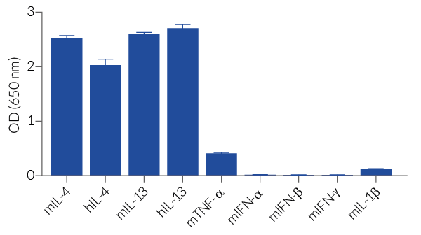
HEK-Blue™ mIL-4/IL-13 cells respond to recombinant mouse IL-4 and recombinant mouse IL-13. They also respond to recombinant human IL-4 and recombinant human IL-13. The reliable and consistent performance of these cells makes them suitable for release assays of molecules that inhibit IL-4 or IL-13 signaling, such as monoclonal antibodies targeting IL-4 or IL-13, or their receptor, and cross-reacting with their human counterparts (see figures).
Key features
- Readily assessable STAT6-SEAP reporter activity
- Convenient readout using QUANTI-Blue™ Solution
- High sensitivity to mouse (m) and human (h) IL-4 and IL-13
- Stability guaranteed for 20 passages
Applications
- Therapeutic development
- Drug screening
- Release assay
IL-4 and IL-13 are cytokines that play an important role in the regulation of inflammation and immune responses.

Disclaimer: These cells are for internal research use only and are covered by a Limited Use License (See Terms and Conditions). Additional rights may be available.
SPECIFICATIONS
Specifications
IL-4, IL-13
Mouse, Human
Detection and quantification of mouse IL-4 or IL-13 activity
0.01 - 1 ng/ml (mIL-4, mIL-13, hIL-4, and hIL-13)
Complete DMEM (see TDS)
Verified using Plasmotest™
Each lot is released only after meeting predefined biological activity specifications in a cell-based assay.
CONTENTS
Contents
-
Product:HEK-Blue™ mIL-4/IL-13 Cells
-
Cat code:hkb-mil413-4
-
Quantity:3-7 x 10^6 cells
- 2 x 1 ml of HEK-Blue™ Selection (250x concentrate)
- 1 ml of Normocin® (50 mg/ml)
- 1 ml of QB reagent and 1 ml of QB buffer (sufficient to prepare 100 ml of QUANTI-Blue™ Solution, a SEAP detection reagent)
Shipping & Storage
- Shipping method: Dry ice
- Liquid nitrogen vapor
- Upon receipt, store immediately in liquid nitrogen vapor. Do not store cell vials at -80°C.
Storage:
Caution:
Details
Cell line description
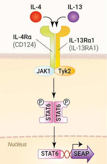
HEK-Blue™ mIL-4/IL-13 cells were generated by stable transfection of the human embryonic kidney HEK293 cell line with genes encoding the mouse IL-4Rα and IL-13Rα1 ((IL-13RA) receptor chains. These cells were further transfected with an STAT6-inducible secreted embryonic alkaline phosphatase (SEAP) reporter. The binding of IL-4 or IL-13 to their receptor triggers a signaling cascade leading to STAT6 activation and the subsequent production of SEAP. This can be readily assessed in the supernatant using QUANTI-Blue™ Solution, a SEAP detection reagent.
HEK-Blue™ mIL-4/IL-13 cells are highly sensitive to mouse (m) IL-4 and IL-13 and also respond to their human counterparts.
IL-4 and IL-13 background
IL-4 and IL-13 are cytokines that belong to the IL-2/γc superfamily. Both cytokines are produced as secreted monomeric molecules by activated CD4+ T cells, Natural Killer T cells (NKT cells), group 2 innate lymphoid cells (ILC2s), macrophages, mast cells, basophils, or eosinophils [1, 2].
IL-4 binds two types of heterodimeric receptors. The type I receptor is composed of the IL-4Rα and the common γ (γc) subunits, while the type II receptor is composed of the IL-4Rα and IL-13Rα1 subunits. IL-13 binds the type II receptor, which is therefore shared with IL-4. The binding of IL-4 or IL-13 to their receptors triggers a signaling cascade leading to the activation of STAT6. Subsequent gene expression drives M2 macrophage polarization, B-cell proliferation, IgE immunoglobulin class switching, and other type 2 immune responses [1, 2]. In non-hematopoietic cells, their functions include mucus production and tissue remodeling. Altogether, the downstream IL-4 and IL-13 effector functions participate in parasitic infection clearance and allergic reactions [1-3].
Relevance for therapeutics development
IL-4 and IL-13 are key cytokines for controlling infections by extracellular parasites. However, they also contribute to harmful allergic responses [2, 3]. As the prevalence of chronic allergic diseases such as asthma and atopic dermatitis is increasing worldwide, there has been a keen interest in the therapeutic blocking of IL-4 and IL-13 signaling.
Dupilumab is a fully human monoclonal antibody (mAb) that targets the IL-4Rα subunit [4]. It acts like a receptor antagonist and inhibits the signaling of both IL-4 and IL-13 [4]. Dupilumab was FDA-approved in 2017 for treating asthma, atopic dermatitis, and chronic sinusitis [4, 5]. Due to its favorable safety profile and established clinical use, Dupilumab holds significant promise for amending treatment options for many dermatologic conditions [6].
Tralokinumab and Lebrikizumab are fully human mAbs that target IL-13, inhibiting its receptor binding ability. Tralokinumab and Lebrikizumab were FDA-approved in 2021 and 2024, respectively, for treating moderate-to-severe atopic dermatitis [7, 8]. The development of engineered IL-4 cytokines has also been explored, including IL-4 "superkines" featuring a higher affinity for type I or type II receptors [9].
References:
1. Keegan, A.D., et al., 2021. Recent advances in understanding the role of IL-4 signaling. Fac Rev. 10: 71.
2. Bernstein, Z.J., et al., 2023. Engineering the IL-4/IL-13 axis for targeted immune modulation. Immunol Rev. 320(1):29-57.
3. Ogulur, I., et al., 2025. Type 2 immunity in allergic diseases. Cell & Mol Immunol, 22(3):211-242.
4. Harb, H. & Chatila, T.A., 2020. Mechanisms of Dupilumab. Clin Exp Allergy. 50(1):5-14.
5. https://www.accessdata.fda.gov/drugsatfda_docs/label/2022/761055s040lbl.pdf
6. Olbrich, H., et al., 2023. Dupilumab in Inflammatory Skin Diseases: A Systematic Review. Biomolecules. 13(4):634.
7. https://www.accessdata.fda.gov/drugsatfda_docs/nda/2022/761180Orig1s000lbl.pdf
8. https://www.accessdata.fda.gov/drugsatfda_docs/label/2024/761306Orig1s000correctedlbl.pdf
9. Leonard, W.J & Lin, J.X., 2023. Strategies to therapeutically modulate cytokine action. Nat Rev Drug Discov. 22(10):827-854.
DOCUMENTS
Documents
Technical Data Sheet
Validation Data Sheet
Safety Data Sheet
Certificate of analysis
Need a CoA ?
You may also need



Frequently Asked Questions
HEK293 cell line description
We presume them to be in the endosome as we express the wild-type, full-length genes. Additionally, their signal can be blocked by Chloroquine, an endosomal acidification inhibitor. However, as these TLRs are over-expressed there may potentially be low expression of them on the cell surface.
HEK293 cells express TLR1, TLR3, TLR5, TLR6, and NOD1.
They respond to TLR3, TLR5, and NOD1 agonists, but at a much lower level compared to HEK293 cells transfected with these receptors
In the United States, HEK293 cell lines are designated Biosafety Level 2 according to the Center for Disease Control and Prevention (CDC).
In Germany, HEK293 cell lines are designated Biosafety Level 1 according to the Central Committee of Biological Safety, Zentrale Kommission für die Biologische Sicherheit (ZKBS).
You can check with your country’s regulatory authority regarding the use of these cells.
Please note that there is no replicating/infectious Adenovirus 5 in these cells.
The minimal promoter isn't the same but the difference in expression for these two Null cell lines is minor.
There is no specific integration system used to generate our stable cell lines.
The selection pressure is enough to obtain stable clones. The receptors are added by simple transfection of plasmids using a cationic lipidic transfection agent (the plasmids are not linearized before transfection).
Only our HEK-Blue™ Null1-k and Null2-k cells are sensitive to G418.
The only HEK-Blue™ Null cells that are sensitive to Puromycin are the HEK-Blue™ Null1-v cells.
The only HEK-Blue™ Null cells that are sensitive to Puromycin are the HEK-Blue™ Null1-v cells.
Our RNAseq data confirms that our HEK-Blue™ cells express FcRN, however, this has not been functionally tested.
The difference in activity is approximately 10-fold.
Both cell types overexpress a designated TLR. The primary difference is that HEK-Blue™ cells include the NFκB inducible SEAP reporter construct.
HEK-Blue™ cells only express a single NFκB inducible SEAP reporter gene. Whereas, HEK-Dual™ cells have the addition of the Lucia™ gene knocked into the IL-8 locus. Thus, when IL-8 is activated following stimulation, HEK-Dual™ cells can report this with the secretion of Lucia™ luciferase.
It should also be noted that the HEK-Dual™ cells have been knocked out for TLR3, TLR5, and TNFR to limit interference from other TLRs when studying a specific TLR pathway.
HEK-Blue™ IL-1R cells express both human and murine IL-1β receptors, thus can detect both species.
On the other hand, HEK-Blue™ IL-1β cells are specific for human IL-1β, but can still detect higher concentrations of mouse IL-1β.
Cell line culture
The split ratio will depend on when you expect confluency. Typically, the doubling time of HEK-Blue™ cells is approximately 24 hours.
Therefore, if you use a split ratio of 1:2 (50%) into a new flask, cells should be confluent the following day. If you use a split ratio of 1:4 (25%) you can expect the cells to be confluent after 2 days.
Our HEK-Blue™ Selection is provided in 1ml tubes with each containing a 250X solution.
Therefore, you should dilute HEK-Blue™ Selection 1:250 into your media to have a 1X concentration.
HEK-Blue™ cells should be seeded at a density of approximately 1.5 x 10⁶ cells in a T25 flask or 4 – 5 x 10⁶ cells in a T75 flask.
We recommend using a flat-bottom, clear walled cell culture plate.
Below are a few tips we recommend to help get your HEK cells growing:
• For the first 2-3 passages, grow cells in media containing 20% FBS and no antibiotics.
• Do not allow cells to reach 100% confluency Please check cells as regularly as possible.
• The cells should not be grown in 20% FBS for too long. Use media with 10% FBS after 2 or 3 passages.
• When making frozen stocks, continue growing additional cultures in case there is a problem with the frozen stock.
Trypsin does not adversely affect the health or growth of these cells. However, it is known that high concentrations will occasionally induce the activation of NFκB resulting in a higher background in your assay.
Moreover, we have observed some cases where trypsin has been contaminated with TLR2, TLR4, and TLR5 contaminants, which can also interfere with the assay results.
It is not unusual for different TLR cells to grow at different rates. Some TLR clones happen to grow a little slower/faster than others. This is often clone dependent.
When the HEK-Blue™ cells are non-adherent, either they were diluted too harshly at the start or they have grown over-confluent in a small flask and suffocated.
To avoid this in the future:
• Change the media and plate the cells at a density of approximately 1.5 x 10⁶ cells in a T25 flask.
• Wash the cells before putting them into a new flask. Sometimes when the cells are non-adherent, it is due to the clustering of both live and dead cells. Additionally, this will get rid of any remaining DMSO which could affect the adhesion of the cells to the flask.
• Use medium with 20% FBS.
• The use of CellBIND flasks can sometimes help to increase attachment and growth of the cells (however CellBIND flasks are not required in the normal protocol).
Assays
There are 2 possible explanations as to why a blue color is observed in all wells.
1. It could be due to the presence of Alkaline Phosphatase (AP) in the culture medium. To see if this is the case, there is a very simple test to perform. Add 50 µl of the medium used for cell culture (without cells) and 200 µl of resuspended HEK-Blue™ detection medium or QUANTI-Blue™. If the medium turns blue, then it is due to the presence of Alkaline Phosphatase (AP) in the serum of the media. In this case, you must heat the serum to inactivate the AP and repeat the medium test. At this point the test should give a negative result (no blue color).
2. It could be due to improper handling of cells before the test. To avoid activation of NFκB before stimulation and reduce the risk of false positive results:
• Use pre-warmed PBS to wash cells
• Use heat-inactivated FBS
• Do not centrifuge cells prior to stimulation
• Do not use trypsin
We have noticed a loss of sensitivity when using HEK-Blue™ Detection medium instead of QUANTI-Blue™ on our cytokine reporter cell lines.
Therefore, we recommend using QUANTI-Blue™, which is provided with the cells, as this is what we use in house.
We recommend to not use any antibiotics at all during assays to ensure the least amount of potential interfering agents in the medium.
Therefore, we do not add HEK-Blue™ Selection to the test media.
We have only tested the use of plasma and serum samples on our HEK-Blue™ hTLR2 cell line.
The results demonstrated that when compared to using standard samples (in DMEM), serum samples give a single log difference.
On the other hand, we found a 3-log difference between DMEM and plasma samples.
This is why we would recommend using serum samples over plasma samples.
Yes, they can be used interchangeably. However, please note that the protocols are distinctly different and need to be followed accordingly.
HEK293 cells are very easy to transfect with a transfection efficiency of approximately 80%.
It depends on the cell line and the concentration of the ligand used to stimulate the cells. In general, we record the results following 16 – 24 hours of stimulation.
 Powered by Bioz