



HEK-Blue™ IL-2Rβ/γ Cells
-
With a free vial of human IL-2 protein
-
Cat.code:
hkb-hil2rbgv2-2NEW
- Documents
ABOUT
IL-2/IL-15 STAT5-SEAP reporter assay in a low-CD122/CD132 receptor background
HEK-Blue™ IL-2Rβ/γ cells are designed to evaluate IL-2- and IL-15-mediated STAT5 activation in a cellular environment expressing low levels of CD122 and CD132. This reporter cell line can be used to assess cytokine bioactivity and the activity of molecules that modulate IL-2- and IL-15-dependent signaling pathways.
HEK-Blue™ IL-2Rβ/γ cells respond to recombinant human IL-15 and, to a lesser extent, human IL-2, while showing no response to their mouse (m) counterparts (see figures). Compared to HEK-Blue™ CD122/CD132 cells, their lower CD122/CD132 expression profile makes them particularly suitable for IL-2 and IL-15 biologic screening under limited receptor availability and for anti-hIL-2 neutralization assays.
Key features
- Readily assessable STAT5-SEAP reporter activity
- Convenient readout using QUANTI-Blue™ Solution
- Detection of human (h) IL-2 and IL-15 within a low CD122/CD132 background
- Suitable for anti-hIL-2 neutralization assays
- Stability guaranteed for 20 passages
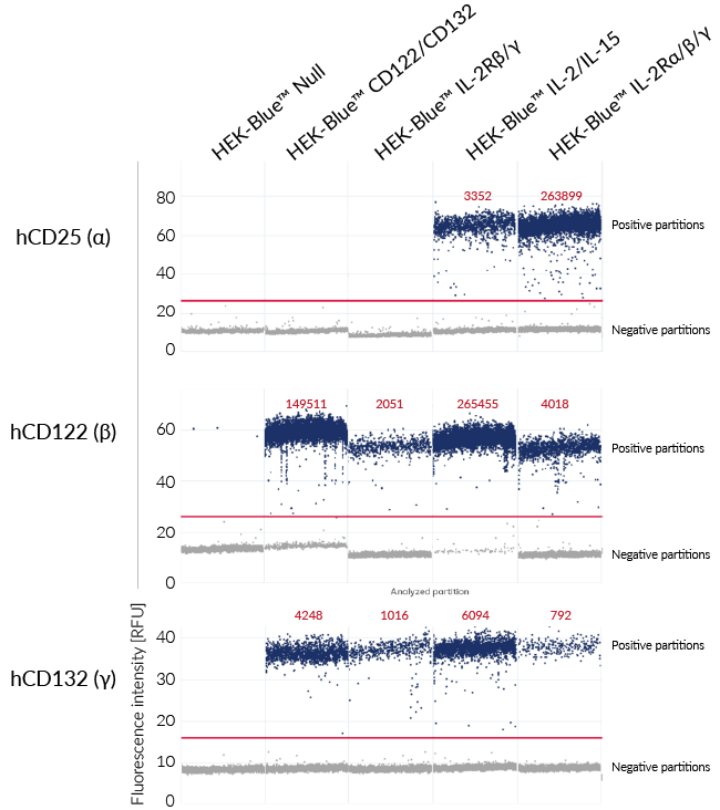
InvivoGen offers four IL-2 and IL-15 reporter cell lines, HEK-Blue™ IL-2Rβ/γ, HEK-Blue™ CD122/CD132, HEK-Blue™ IL-2Rα/β/γ, and HEK-Blue™ IL-2/IL-15, with distinct receptor expression profiles and cytokine detection potencies (see table below). Digital PCR was used to confirm the expression profiles of these three IL-2 receptor subunits in the HEK-Blue™ reporter cell lines (see image carousel above).
| Reporter cell line | Receptor expression | Cytokine detection potency | |||||
|---|---|---|---|---|---|---|---|
hCD25 (α) | hCD122 (β) | hCD132 (γ) | hIL-2 | hIL-15 | mIL-2 | mIL-15 | |
| HEK-Blue™ IL-2Rα/β/γ | High | Low | Low | ++++ | +++ | ++ | - |
| HEK-Blue™ IL-2/IL-15 Cells | Low | High | High | +++ | ++++ | ++ | - |
| HEK-Blue™ IL-2Rβ/γ | None | Low | Low | + | ++ | - | - |
| HEK-Blue™ CD122/CD132 | None | High | High | ++++ | ++++ | +/- | +/- |
IL-2 and IL-15 are closely related cytokines that belong to the IL-2 family and display important functions in the immune system . They share the heterodimeric CD122/CD132 receptor to deliver their signals within target cells. Their specificity of action is conferred by their α receptor chains, CD25 and CD215 (IL-15Rα).

Disclaimer: These cells are for internal research use only and are covered by a Limited Use License (See Terms and Conditions). Additional rights may be available.

SPECIFICATIONS
Specifications
IL-2
IL-15
Human
0.3 - 3 ng/ml (hIL-2)
0.1 - 1 ng/ml (hIL-15)
Complete DMEM (see TDS)
Verified using Plasmotest™
Each lot is functionally tested and validated.
CONTENTS
Contents
-
Product:HEK-Blue™ IL-2Rβ/γ Cells
-
Cat code:hkb-hil2rbgv2-2
-
Quantity:3-7 x 10^6 cells
- 2 x 1 ml of HEK-Blue™ Selection (250x concentrate)
- 1 ml Normocin® (50 mg/ml)
- 1 ml of QB reagent and 1 ml of QB buffer (sufficient to prepare 100 ml of QUANTI-Blue™ Solution, a SEAP detection reagent).
Shipping & Storage
- Shipping method: Dry ice
- Liquid nitrogen vapor
- Upon receipt, store immediately in liquid nitrogen vapor. Do not store cell vials at -80°C.
Storage:
Caution:
Details
Cell line description
HEK-Blue™ IL-2Rβ/γ cells were generated by stable transfection of the human embryonic kidney HEK293 cell line with the genes encoding the human CD122 (IL-2Rβ) and CD132 (IL-2Rγ) subunits. These cells were engineered to express low levels of CD122 and CD132, enabling the detection of low-affinity IL-2/IL-15 signaling independently of CD25 (IL-2Rα). They also feature stable expression of human JAK3 and STAT5 to obtain a functional IL-2/IL-15 signaling pathway, as well as a STAT5-inducible secreted embryonic alkaline phosphatase (SEAP) reporter. The binding of IL-2 or IL-15 to their receptors triggers a signaling cascade leading to STAT5 activation and subsequent SEAP production. This can be readily assessed in the culture supernatant using QUANTI-Blue™ Solution, a SEAP detection reagent.
HEK-Blue™ IL-2Rβ/γ cells respond to both human IL-2 and IL-15, with greater responsiveness to IL-15 than IL-2. Their low CD122/CD132 expression profile and lack of CD25 expression make them particularly useful for evaluating IL-2 and IL-15 biologics in a low-receptor environment and for assessing molecules that modulate signaling through the shared CD122/CD132 receptor complex. In addition, these cells can be used to validate the functionality, potency, toxicity, and dose-dependent effects of IL-2- and IL-15-based biologics.
IL-2 and IL-15 background
Interleukin-2 (IL-2) and interleukin-15 (IL-15) are closely related cytokines that play central roles in the regulation of innate and adaptive immune responses [1,2]. Both cytokines signal through receptor complexes containing the CD122 and CD132 subunits, while their biological specificity is conferred by distinct α receptor chains: CD25 (IL-2Rα) for IL-2 and CD215 (IL-15Rα) for IL-15 [1,3]. The incorporation of CD25 into the receptor complex generates the highest-affinity IL-2 receptor. This enables cells expressing elevated CD25 levels to respond efficiently to low concentrations of IL-2 [3,4].
The binding of IL-15 and IL-2 to their heterodimeric or heterotrimeric receptors activates JAK1/JAK3-STAT5 signaling together with the PI3K/AKT/mTOR and MAPK pathways, resulting in cellular responses such as proliferation, survival, differentiation, and immune effector functions [1,3]. IL-2 is a key regulator of activated T cells and regulatory T cells (Tregs), whereas IL-15 plays an important role in the development, maintenance, and activation of natural killer (NK) cells and memory CD8+ T cells [1,2].
Aberrant IL-2 and IL-15 signaling has been associated with T-cell large granular lymphocytic leukemia, cutaneous T-cell lymphoma, adult T-cell leukemia/lymphoma, multiple myeloma, celiac disease, rheumatoid arthritis, psoriasis, and inflammatory bowel disease, while CD25 overexpression has been reported in several hematological malignancies and solid tumors [1-4]. Consequently, IL-2-, IL-15-, and CD25-directed therapies are being actively developed for the treatment of cancer, autoimmune disorders, and chronic inflammatory diseases [1-4].
1. Sindaco P, et al., 2023. The role of interleukin-15 in the development and treatment of hematological malignancies. Front Immunol. 14:1141208
2. Propper DJ & Balkwill FR, 2022. Harnessing cytokines and chemokines for cancer therapy. Nat. Rev. Clin. Oncol. 19:237-253.
3. Peng Y, et al., 2022. CD25: A potential tumor therapeutic target. Int J Cancer. 151:1617-1626.
4. Al Nasar M, et al., 2024. The T-Cell Growth Factor Interleukin-2, Which Is Occasionally Targeted by Autoantibodies, Qualifies as Drug for the Treatment of Allergy, Autoimmunity, and Cancer: Collegium Internationale Allergologicum (CIA) Update 2024. Int Arch Allergy Immunol. 185:286-300.
DOCUMENTS
Documents
Technical Data Sheet
Validation Data Sheet
Safety Data Sheet
Certificate of analysis
Need a CoA ?
You may also need



Frequently Asked Questions
HEK293 cell line description
We presume them to be in the endosome as we express the wild-type, full-length genes. Additionally, their signal can be blocked by Chloroquine, an endosomal acidification inhibitor. However, as these TLRs are over-expressed there may potentially be low expression of them on the cell surface.
HEK293 cells express TLR1, TLR3, TLR5, TLR6, and NOD1.
They respond to TLR3, TLR5, and NOD1 agonists, but at a much lower level compared to HEK293 cells transfected with these receptors
In the United States, HEK293 cell lines are designated Biosafety Level 2 according to the Center for Disease Control and Prevention (CDC).
In Germany, HEK293 cell lines are designated Biosafety Level 1 according to the Central Committee of Biological Safety, Zentrale Kommission für die Biologische Sicherheit (ZKBS).
You can check with your country’s regulatory authority regarding the use of these cells.
Please note that there is no replicating/infectious Adenovirus 5 in these cells.
The minimal promoter isn't the same but the difference in expression for these two Null cell lines is minor.
There is no specific integration system used to generate our stable cell lines.
The selection pressure is enough to obtain stable clones. The receptors are added by simple transfection of plasmids using a cationic lipidic transfection agent (the plasmids are not linearized before transfection).
Only our HEK-Blue™ Null1-k and Null2-k cells are sensitive to G418.
The only HEK-Blue™ Null cells that are sensitive to Puromycin are the HEK-Blue™ Null1-v cells.
The only HEK-Blue™ Null cells that are sensitive to Puromycin are the HEK-Blue™ Null1-v cells.
Our RNAseq data confirms that our HEK-Blue™ cells express FcRN, however, this has not been functionally tested.
The difference in activity is approximately 10-fold.
Both cell types overexpress a designated TLR. The primary difference is that HEK-Blue™ cells include the NFκB inducible SEAP reporter construct.
HEK-Blue™ cells only express a single NFκB inducible SEAP reporter gene. Whereas, HEK-Dual™ cells have the addition of the Lucia™ gene knocked into the IL-8 locus. Thus, when IL-8 is activated following stimulation, HEK-Dual™ cells can report this with the secretion of Lucia™ luciferase.
It should also be noted that the HEK-Dual™ cells have been knocked out for TLR3, TLR5, and TNFR to limit interference from other TLRs when studying a specific TLR pathway.
HEK-Blue™ IL-1R cells express both human and murine IL-1β receptors, thus can detect both species.
On the other hand, HEK-Blue™ IL-1β cells are specific for human IL-1β, but can still detect higher concentrations of mouse IL-1β.
Cell line culture
The split ratio will depend on when you expect confluency. Typically, the doubling time of HEK-Blue™ cells is approximately 24 hours.
Therefore, if you use a split ratio of 1:2 (50%) into a new flask, cells should be confluent the following day. If you use a split ratio of 1:4 (25%) you can expect the cells to be confluent after 2 days.
Our HEK-Blue™ Selection is provided in 1ml tubes with each containing a 250X solution.
Therefore, you should dilute HEK-Blue™ Selection 1:250 into your media to have a 1X concentration.
HEK-Blue™ cells should be seeded at a density of approximately 1.5 x 10⁶ cells in a T25 flask or 4 – 5 x 10⁶ cells in a T75 flask.
We recommend using a flat-bottom, clear walled cell culture plate.
Below are a few tips we recommend to help get your HEK cells growing:
• For the first 2-3 passages, grow cells in media containing 20% FBS and no antibiotics.
• Do not allow cells to reach 100% confluency Please check cells as regularly as possible.
• The cells should not be grown in 20% FBS for too long. Use media with 10% FBS after 2 or 3 passages.
• When making frozen stocks, continue growing additional cultures in case there is a problem with the frozen stock.
Trypsin does not adversely affect the health or growth of these cells. However, it is known that high concentrations will occasionally induce the activation of NFκB resulting in a higher background in your assay.
Moreover, we have observed some cases where trypsin has been contaminated with TLR2, TLR4, and TLR5 contaminants, which can also interfere with the assay results.
It is not unusual for different TLR cells to grow at different rates. Some TLR clones happen to grow a little slower/faster than others. This is often clone dependent.
When the HEK-Blue™ cells are non-adherent, either they were diluted too harshly at the start or they have grown over-confluent in a small flask and suffocated.
To avoid this in the future:
• Change the media and plate the cells at a density of approximately 1.5 x 10⁶ cells in a T25 flask.
• Wash the cells before putting them into a new flask. Sometimes when the cells are non-adherent, it is due to the clustering of both live and dead cells. Additionally, this will get rid of any remaining DMSO which could affect the adhesion of the cells to the flask.
• Use medium with 20% FBS.
• The use of CellBIND flasks can sometimes help to increase attachment and growth of the cells (however CellBIND flasks are not required in the normal protocol).
Assays
There are 2 possible explanations as to why a blue color is observed in all wells.
1. It could be due to the presence of Alkaline Phosphatase (AP) in the culture medium. To see if this is the case, there is a very simple test to perform. Add 50 µl of the medium used for cell culture (without cells) and 200 µl of resuspended HEK-Blue™ detection medium or QUANTI-Blue™. If the medium turns blue, then it is due to the presence of Alkaline Phosphatase (AP) in the serum of the media. In this case, you must heat the serum to inactivate the AP and repeat the medium test. At this point the test should give a negative result (no blue color).
2. It could be due to improper handling of cells before the test. To avoid activation of NFκB before stimulation and reduce the risk of false positive results:
• Use pre-warmed PBS to wash cells
• Use heat-inactivated FBS
• Do not centrifuge cells prior to stimulation
• Do not use trypsin
We have noticed a loss of sensitivity when using HEK-Blue™ Detection medium instead of QUANTI-Blue™ on our cytokine reporter cell lines.
Therefore, we recommend using QUANTI-Blue™, which is provided with the cells, as this is what we use in house.
We recommend to not use any antibiotics at all during assays to ensure the least amount of potential interfering agents in the medium.
Therefore, we do not add HEK-Blue™ Selection to the test media.
We have only tested the use of plasma and serum samples on our HEK-Blue™ hTLR2 cell line.
The results demonstrated that when compared to using standard samples (in DMEM), serum samples give a single log difference.
On the other hand, we found a 3-log difference between DMEM and plasma samples.
This is why we would recommend using serum samples over plasma samples.
Yes, they can be used interchangeably. However, please note that the protocols are distinctly different and need to be followed accordingly.
HEK293 cells are very easy to transfect with a transfection efficiency of approximately 80%.
It depends on the cell line and the concentration of the ligand used to stimulate the cells. In general, we record the results following 16 – 24 hours of stimulation.
 Powered by Bioz