
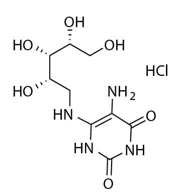
5-A-RU
-
Cat.code:
tlrl-5aruNEW
- Documents
ABOUT
5-A-RU with hydrochloride - MAIT activator precursor - CAS# 134452-11-2
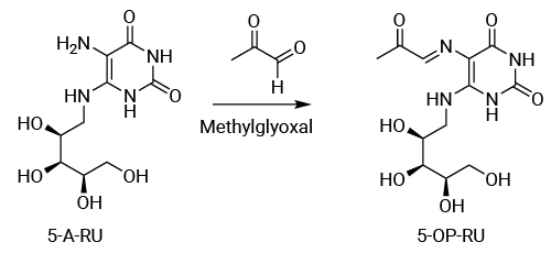
5-A-RU (5-Amino-6-(D-ribitylamino)uracil) is an intermediate of the microbial riboflavin (vitamin B2) biosynthetic pathway. 5-A-RU is a precursor of potent MR1 ligands to activate mucosal-associated invariant T (MAIT) cells. In combination with methylglyoxal, 5-A-RU enables the in situ generation of 5-OP-RU, a potent MAIT cell agonist presented by the MHC class I-related protein 1 (MR1) on various target cells.

5-A-RU is inherently unstable and can rapidly degrade following preparation. InvivoGen's 5-A-RU is provided as a hydrochloride salt (5-A-RU·HCl), a stabilized form. Conversion to its hydrochloride salt, 5-A-RU·HCl improves its stability and facilitates its storage and handling, without interfering with biological functionality and further MR1 ligand generation.
Key features
- Lot-to-lot validated bioactivity using a functional MAIT cell assay
- Supplied with hydrochloride for improved stability
- Endotoxin tested
Applications
- MAIT cell activation assays
- MR1 antigen presentation studies
- MR1/5-OP-RU tetramer generation
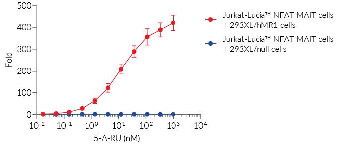
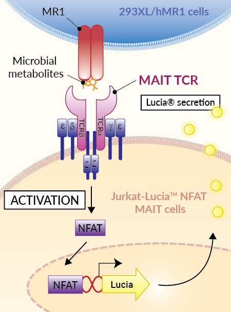
The biological activity of 5-A-RU can be assessed using InvivoGen's co-culture system of MR1-exressing HEK293 target cells and Jurkat-Lucia™ NFAT MAIT reporter cells. In the presence of methylglyoxal (not provided), 5-A-RU generates 5-OP-RU, which is presented by MR1 on the target cells. Recognition of the MR1–5-OP-RU complex by the MAIT TCR activates NFAT signaling and induces secretion of Lucia® luciferase, providing a quantitative readout of MR1-dependent MAIT TCR activation. Lucia® activity in the supernatant is readily quantified using QUANTI-Luc™ 4 Lucia/Gaussia (see figure).
InvivoGen's 5-A-RU provides a convenient research tool to activate MAIT cells, investigate MR1-dependent antigen presentation, study MAIT TCR signaling, and develop functional MAIT cell assays. It can be used with MR1-expressing antigen-presenting cells, primary immune cells, or MAIT reporter cell systems to investigate the MR1-MAIT axis.
All InvivoGen products are for internal research use only, and not for human or veterinary use.
SPECIFICATIONS
Specifications
MR1
C9H16N4O6 ·HCl
1 pM – 1 µM
Cellular assay
Each lot is released only after meeting predefined biological activity specifications in a cell-based assay.
The lyophilized product is very lightweight and may become dispersed during handling. Before opening the vial, briefly centrifuge it to ensure that all the lyophilized material is collected at the bottom of the vial.
CONTENTS
Contents
-
Product:5-A-RU
-
Cat code:tlrl-5aru
-
Quantity:1 mg
Shipping & Storage
- Shipping method: Room temperature
- -20°C
- Avoid repeated freeze-thaw cycles
- Protect from light
Storage:
Caution:
Details
What are MAIT cells?
Mucosal-associated invariant T (MAIT) cells represent a conserved arm of the immune system, bridging the innate and adaptive responses1. They are one of the most abundant populations of unconventional T cells in humans, accounting for 1% to 10% of circulating T cells. They are especially enriched in barrier and mucosal tissues, as well as in the liver (up to 40% of resident T cells), lung, and intestine, where they play a key role in antimicrobial defense1-2. MAIT cells express specialized semi-invariant T cell antigen receptors (TCRs), which recognize small metabolite antigens presented by the non-polymorphic MHC class I-related protein (MR1)3.
What is MR1?
MR1 (MHC class I-related protein 1) is an antigen-presenting molecule that presents small metabolite antigens to MAIT cells and other MR1-restricted T cells. MR1 is a highly conserved membrane protein among mammalian species and associates non-covalently with β2-microglobulin (β2m). Although MR1 is broadly transcribed, its cell-surface expression is normally limited. Under physiological conditions, MR1 is localized in the endoplasmic reticulum. During infection, MR1 molecules associated with suitable ligands, such as microbial vitamin B metabolites, migrate to the cell surface, and present them to and, thus, activate MAIT cells3.
What is 5-A-RU?
5-Amino-6-D-ribitylaminouracil (5-A-RU) is an intermediate of the microbial riboflavin (vitamin B2) biosynthetic pathway and a precursor of potent MR1-presented MAIT cell antigens. Many bacteria and fungi, but not mammalian cells, possess the riboflavin biosynthetic pathway and can generate 5-A-RU3. Importantly, 5-A-RU does not directly bind MR1 or activate MAIT cells. Instead, it reacts non-enzymatically with small carbonyl metabolites that can originate from host or microbial metabolism. Reaction with methylglyoxal, an abundant by-product of glycolysis, generates the potent but unstable MAIT agonist 5-OP-RU, whereas reaction with glyoxal generates 5-OE-RU4,5. 5-OP-RU is captured within the MR1 antigen-binding cleft, where it forms a covalent Schiff base with Lys43 (K43) of MR14. This interaction stabilizes the MR1–5-OP-RU complex and promotes its trafficking to the cell surface for recognition by the MAIT TCR4. Because 5-OP-RU is unstable in aqueous solution, it is commonly generated in situ from 5-A-RU and methylglyoxal for the study of MR1-dependent MAIT cell activation5.
How are MAIT cells activated?
MAIT cells are able to sense a wide range of bacteria and can be rapidly activated and mount potent immune responses through two distinct pathways1,3.
In the TCR-dependent pathway, microbial metabolites are loaded and presented by the MR1 molecule on the surface of infected cells, leading to MAIT cell activation. Alternatively, MAIT cells can be triggered in an MR1-independent manner through inflammatory cytokines, notably IL-12 and IL-18, released during infection1.
As a result, MAIT cells secrete a variety of pro-inflammatory cytokines and chemokines (e. g. IFN-γ, TNF, IL-17), as well as express the cytotoxic molecules, granzyme B and perforin. With their expression of immune checkpoint receptors, strong cytotoxic capabilities, and graft-versus-host-disease (GvHD)-free safety profile, MAIT cells have become an attractive focus in tumor immunology research1.
How are MAIT cells being explored for cancer immunotherapy?
Emerging strategies include MAIT-cell engagers6, such as bispecific antibodies that redirect MAIT cells toward tumor-associated antigens, as well as CAR-engineered MAIT cells for adoptive cell therapy1. Additional approaches aim to expand MAIT cells ex vivo, generate MAIT cells from induced pluripotent stem cells, or enhance their activity through MR1 ligands, cytokines, and microbiota modulation1. Together, these strategies highlight the growing interest in harnessing MAIT cells for cancer immunotherapy and other immune-mediated diseases. However, most MAIT cell-based therapies remain in the preclinical or early clinical stages, and further research is needed to better understand MAIT cell biology and fully realize their therapeutic potential.
References:
1. Yigit M, et al., 2024. Mucosal-associated invariant T cells in cancer: dual roles, complex interactions and therapeutic potential. Front Immunol. 15:1369236.
2. Ghazarian L, et al., 2017. Mucosal-Associated Invariant T Cell Interactions with Commensal and Pathogenic Bacteria: Potential Role in Antimicrobial Immunity in the Child. Front Immunol. 8:1837.
3. Howson LJ, et al., 2015. MR1-Restricted Mucosal-Associated Invariant T Cells and Their Activation during Infectious Diseases. Front Immunol. 6:303.
4. Corbett AJ, et al., 2014. T-cell activation by transitory neo-antigens derived from distinct microbial pathways. Nature. 509:361–365.
5. Li K, et al., 2018. Synthesis, stabilization, and characterization of the MR1 ligand precursor 5-amino-6-D-ribitylaminouracil (5-A-RU). PLoS ONE. 13:e0191837.
6. Plyte SE, et al., 2024. MAIT engagers: Bispecific antibody-mediated redirection of mucosal associated invariant T cells to treat solid tumors. Cancer Res. 84(6_Suppl) 6708.
DOCUMENTS
Documents
Technical Data Sheet
Validation Data Sheet
Safety Data Sheet
Certificate of analysis
Need a CoA ?

 Powered by Bioz