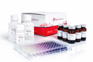
Cell death quantification is central to a broad range of in vitro applications, including immune effector function studies and drug-induced cytotoxicity. Lactate dehydrogenase (LDH) release assays are widely used to measure loss of membrane integrity and provide a simple and scalable approach for assessing cytotoxicity across diverse experimental settings. This review examines the principles and limitations of LDH-based assays, and how refining the colorimetric readout improves measurement reliability.
LDH release as a marker of cytotoxicity
Overview of cytotoxicity measurement approaches
Cytotoxicity encompasses a spectrum of cellular responses to damaging stimuli, ranging from transient alterations in cellular function to irreversible membrane damage and cell death. Historically, a variety of methods have been used to quantify cell death in vitro. Chromium (Cr51) release assays provide cell death quantification with high sensitivity but involve ionizing radiation hazards and regulatory constraints1. Fluorescent approaches based on membrane integrity— such as membrane impermeant DNA dyes (ie. propidium iodide), or amine-reactive viability dyes— enable sensitive single-cell analysis but require specialized instrumentation and multi-step workflows, which can limit their scalability2. These constraints have driven the adoption of colorimetric LDH release assays as practical alternatives for routine cytotoxicity assessment1,2.
LDH release in response to membrane damage
LDH is a stable, cytoplasmic enzyme involved in the lactate metabolic cycle. Upon membrane rupture, LDH is rapidly released into the extracellular environment, where its activity can be assessed to measure cell death modalities such as necrosis, necroptosis, or pyroptosis. LDH release may also occur at late stages of apoptosis, ferroptosis, or oxidative cell death, once the plasma membrane is disrupted. LDH activity measurements therefore primarily report the loss of membrane integrity, rather than the mechanism of cell death.
Colorimetric LDH assay principle
LDH activity is measured through an enzymatic reaction in which LDH oxidizes lactate to pyruvate, concomitantly reducing NAD+ to NADH. NADH subsequently drives the reduction of tetrazolium salts via a catalyst (e.g., diaphorase or phenazine derivative electron mediators), resulting in the formation of a colored (traditionally red) formazan product which can be quantified spectrophotometrically1-4. The formazan signal is directly proportional to extracellular LDH activity and provides a quantitative and linear measure of cell membrane disruption (Fig. 1).
Limitations of conventional LDH assays
Despite their widespread use, conventional LDH assays based on red formazan present several limitations that can impact data interpretation3. These include:
- spectral overlap with phenol red (present in most culture media), leading to elevated background signal,
- high baseline signal from spontaneous LDH release, which can cover up low-level cytotoxicity,
- low dynamic range due to elevated background signal and high baseline signal,
- loss of assay linearity in high-density culture conditions.
Collectively, these issues underline the need for improved LDH assay designs that enhance signal discrimination.
LDH-Blue™ Plus: overcoming key limitations
Refining the colorimetric readout
One approach to overcoming the assay limitations lies in re-engineering the chromogenic output. By shifting the reaction product toward a blue formazan, rather than a red formazan, signal generation can be decoupled from background absorbance contributed by phenol red in the culture medium. We assessed the naked-eye impact of the spectral shift by comparing blue (LDH-Blue™ Plus, InvivoGen) or red (competitors) formazan readouts in a cell lysis assay. In contrast to conventional red formazan signal that visually blends with the culture medium,the blue formazan signal facilitates naked-eye monitoring of the reaction progression and optimal stopping time (Fig. 2). This distinct blue/purple color change improves usability and reduces the risk of signal saturation.
Extending the detection range
We evaluated how the spectral shift affects the detection range of released LDH, defined as the difference between the maximal and minimal absorbance signals. The maximal absorbance signal is obtained by full cell lysis. The minimal absorbance signal comprises the background absorbance (signal from phenol red and LDH from serum added to the culture medium) and the baseline absorbance (spontaneous LDH release by the test cells). Cell lysis detection range in RPMI culture medium is limited when using conventional LDH assays as they can detect background signal from phenol red. In contrast, LDH-Blue™ Plus avoids background signal from phenol red, allowing clearer differentiation between low and high levels of cytotoxicity (Fig. 3).
These data show that using blue formazan improves the assay sensitivity, which is crucial in applications such as early-stage drug screening or low cytotoxicity monitoring. Moreover, there is no need for adapting cell culture conditions.
Ensuring linearity
Accurate cytotoxicity quantification based on LDH release relies on the assumption that formazan production is proportional to the degree of cell membrane disruption. To evaluate this relationship, we assessed the correlation between LDH signal and cell density across multiple cell types, encompassing both adherent and suspension models. Conventional LDH assays exhibit deviations from linearity at higher signal intensities, likely due to increased contributions from background and baseline absorbance. In contrast, LDH-Blue™ Plus maintains a strong linear correlation between cell density and LDH activity (Fig. 4). By minimizing background contribution and preserving signal proportionality, the blue formazan readout enables consistent and reliable quantification across diverse experimental conditions. This reduces the need for strict optimization of cell density prior to performing the assay.
Improving the measure accuracy
Pyroptotic cell death is a major outcome of inflammasome activation, and it is commonly assessed by quantification of LDH release. We compared the percent cytotoxicity in an NLRP3 inflammasome activation setting using LDH-Blue™ Plus and competitor assays. We used THP-1 human monocytes, a well-established cellular model for inflammasome studies, at a seeding density of 2 x 10⁵ cells per well in 96-well plate. When assessed with blue or conventional red formazan readouts, cells treated with the NLRP3 activator nigericin release LDH in the supernatant in a similar dose-dependent manner. However, the LDH activity reported by the conventional red assays appears systematically higher than with the LDH-Blue™ Plus assay (Fig. 5). Importantly, the signal is no longer linear at this cell density for the conventional assays (Fig. 4A).
Outside of the linearity range, the accuracy of pyroptosis measurements is affected, leading to cell death overestimation. This bias can interfere with proper data interpretation, especially when evaluating drug compound toxicity. Therefore, we examined the accuracy of blue and red formazan-based LDH release assays, compared to a flow cytometry reference method measuring membrane integrity with an amine-reactive dye (Live/Dead® assay). In a late-stage cytotoxicity setting (staurosporine-induced apoptosis), conventional LDH release assays report higher apparent cytotoxicity (+21% and 41%) relative to the reference assay, whereas LDH-Blue™ Plus remains closely aligned (+1%) (Fig. 6).
Overall, these findings indicate that elevated background and baseline absorbances in conventional LDH assays introduce a systematic error that inflates apparent cytotoxicity, notably in conditions where membrane damage is limited or occurs at early stages. LDH-Blue™ Plus minimizes overestimation and provides cytotoxicity measures matching a reference assay.
Supporting reproducibility
Reagent stability is a key determinant of assay reproducibility. LDH-Blue™ Plus maintains consistent signal intensity and dynamic range after one month storage at 4°C, while preserving linearity. In contrast, conventional LDH assays show reduced signal intensity and loss of dynamic range following storage, indicative of decreased reagent performance (Fig. 7). By ensuring stable signal generation over time when reagent is stored at 4°C, LDH-Blue™ Plus avoids variability between experiments and supports reliable quantification in routine workflows.
Conclusion
LDH release assays are widely used to quantify cytotoxicity, but conventional assays relying on red formazan production have limitations that can affect measurement reliability and require extensive optimization. LDH-Blue™ Plus is based on a blue formazan colorimetric readout. Shifting from red to blue formazan improves signal discrimination and reduces background interference from phenol red in culture media, resulting in an enhanced detection range (sensitivity) and improved signal linearity (accuracy). Consistent linearity facilitates precise comparison of data obtained from independent experiments and different laboratories. LDH-Blue™ Plus also offers several practical advantages for routine workflows, including clear naked-eye visualization for reaction monitoring and optimal stopping time, compatibility with standard phenol red–containing media, and robust quantification across a wide range of cell densities, thereby reducing optimization requirements. Furthermore, the kit was engineered for improved reagent stability during storage at 4°C. Altogether, these features make LDH-Blue™ Plus a reliable and user-friendly solution for accurate cytotoxicity assessment in research and screening applications.
References
1. Korzeniewski, C. & Callewaert, D. M., 1983. J Immunol Methods. 64(3):313-320.
2. Cummings, B. S. & Schnellmann, R. G., 2004. Curr Protoc Pharmacol. 12(12.8).
3. Kaja, S. et al., 2017. Curr Protoc Toxicol. 72:2.26.1-2.26.10.
4. Decker, T. & Lohmann-Matthes, M. L., 1988. J Immunol Methods. 115(1):61-9.