

Anti-hCTLA4-hIgG1fut
-
Cat.code:
hctla4-mab13
- Documents
ABOUT
Anti-human CLTA4 antibody - Human IgG1 non-fucosylated (high effector functions)
Anti-hCTLA4-hIgG1fut is a Ipilimumab-based research antibody with a non-fucosylated (fut) human IgG1 isotype. Ipilimumab is a fully human IgG1 monoclonal antibody that targets CTLA-4 (also known as CD152), a negative regulator of T cell activation. Ipilimumab is FDA-approved for the treatment of unresectable or metastatic melanoma.

Anti-hCTLA4-hIgG1fut features the variable region of Ipilimumab and a non-fucosylated IgG1 constant region for very high effector functions. For applications requiring high Fc effector activity, InvivoGen also offer Anti-hCTLA4-hIgG1, an Ipilimumab biosimilar research antibody.
Anti-hCTLA4-hIgG1fut is suitable for effector function analysis, screening assays, and cytotoxic bio-assays. It can be used in antibody-dependent cell-mediated cytotoxicity (ADCC) effector activity assays in combination with Jurkat-Lucia™ NFAT-CD16 effector cells and Raji target cells (see figure).
Key features
- Each lot is functionally tested and validated
- The complete sequence of the antibody construct has been verified
- Absence of endotoxins determined by the EndotoxDetect™ assay
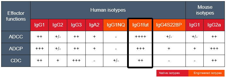
The absence of the fucose residue from the N-glycans of IgG-Fc results in dramatic enhancement of antibody-dependent cellular cytotoxicity (ADCC) without any detectable change in complement-dependent cytotoxicity (CDC) or antigen-binding capability [1,2]. More isotypes of this antibody are available and can be used for comparison of biological activities such as ADCC (see below or in the 'upon request' section).
References:
1. Yamane-Ohnuki N. & Satoh M., 2009. Production of therapeutic antibodies with controlled fucosylation.corresponding MAbs. 1(3): 230–236.
2. Mizushima T., 2011. Structural basis for improved efficacy of therapeutic antibodies on defucosylation of their Fc glycans. Genes Cells. 16(11): 1071–1080.
All products are for internal research use only, and not for human or veterinary use.
SPECIFICATIONS
Specifications
CTLA-4 (CD152, CTLA4)
Human
Neutralization assay, flow cytometry, ELISA, Fc interaction studies
Sodium phosphate buffer with glycine, saccharose and stabilizing agents
Flow cytometry
Each lot is functionally tested and validated.
CONTENTS
Contents
-
Product:Anti-hCTLA4-hIgG1fut
-
Cat code:hctla4-mab13
-
Quantity:100 µg
Shipping & Storage
- Shipping method: Room temperature
- -20°C
- Avoid repeated freeze-thaw cycles
Storage:
Caution:
Details
The cytotoxic T-lymphocyte-associated protein 4 (CTLA-4, CD152) is an inhibitory receptor and immune checkpoint expressed by activated and regulatory T cells [1, 2].
The current paradigm is that full activation of T cells requires at least 2 signals upon contact with antigen-presenting cells (APCs) [3, 4]. Signal 1 is delivered upon the interaction of the T cell receptor (TCR) with antigenic peptides bound to major histocompatibility complex (MHC) molecules on antigen-presenting cells (APCs). Signal 2 is delivered upon the interaction of the co-stimulatory receptor CD28 with the B7 family ligands, B7-1 (CD80) and B7-2 (CD86), on APCs.
Signal 1: TCR and [HLA::peptide]
The 'classical' and most represented TCR is an 80 to 90 kDa heterodimer composed of one α chain and one β chain. The αβTCR is a transmembrane protein expressed by developing and mature T cells. It features an extracellular ligand-binding pocket and a short cytoplasmic tail. Each αβTCR is restricted to a specific complex made of an antigenic peptide and a class I or class II MHC molecule. Human MHC molecules are also known as HLA (human leukocyte antigen). Because of its short cytoplasmic tail, the TCR, once engaged, cannot signal and requires non-covalent association with the CD3 to trigger downstream intracellular signaling and T cell activation [3, 4]. Importantly, signal 1 without co-stimulation results in T cell unresponsiveness or 'anergy', a tolerance mechanism that guards against premature activation.
Signal 2: CD28 and CD80/86
CD28 is a homodimeric and transmembrane protein expressed by T cells. Nearly all human CD4+ T cells and 50% of human CD8+ T cells express CD28. The CD28 interaction with CD80 (aka B7-1) or CD86 (aka B7-2) on APCs, in conjunction with TCR engagement, triggers a co-stimulation signal (signal 2). It results in T-cell proliferation, cytokine production, cell survival, and cellular metabolism [3, 4].
IC signal: CTLA-4 and CD80/86
CTLA-4 exerts competitive binding to the co-stimulatory receptor CD28 ligands (i.e. CD80 and CD86) expressed by antigen-presenting cells. Thereby CTLA-4 upregulation by T cells prevents overstimulation.
Anti-CTLA-4 monoclonal antibodies (mAbs), as well as other immune checkpoints targeting mAbs, are extensively investigated to treat various cancers [2, 5, 6].
Ipilimumab background
Ipilimumab, a fully human IgG1 monoclonal antibody targets the Cytotoxic T-lymphocyte Associated Protein 4 (CTLA-4), also known as CTLA4 or CD152. CTLA-4 is an important immune checkpoint and negative regulator of T cell activation. Upon binding to CTLA-4, Ipilimumab inhibits negative signals that physiologically downregulate T cell activation and exerts its therapeutic activity by upregulating the antitumor activity of T lymphocytes [7,8]. In addition, Ipilimumab induces antibody-dependent cell-mediated cytotoxicity (ADCC) and TNF-α production [9]. Ipilimumab is FDA-approved for the treatment of unresectable or metastatic melanoma and is undergoing various clinical trials for other types of cancers, including lung cancer [10].
References:
1. Ribas A. and Wolchock J.D. 2018. Cancer immunotherapy using checkpoint blockade. Science. 359:1350.
2. Wei, S.C. et al. 2018. Fundamental mechanisms of immune checkpoint blockade therapy. Cancer Discov. 8(9):1069.
3. Budd R.C. & Fortner K.A., 2017. Chapter 12 - T Lymphocytes. Kelley and Firestein's Textbook of Rheumatology (Tenth Edition). pages 189-206.
4. Smith-Garvin J.E. et al., 2009. T Cell Activation. Ann. Rev. Immunol. 27:591-619.
5. Wilson, R.A.M. et al. 2018. Immune checkpoint inhibitors: new strategies to checkmate cancer. Clin. Exp. Immunol. 191(2):133-148.
6. Marin-Acevedo J.A. et al. 2018. Next generation of immune checkpoint therapy in cancer: new developments and challenges. J. Hematol. Oncol. 11(1):39.
7. Grosso JF. & Jure-Kunkel MN., 2013. CTLA-4 blockade in tumor models: an overview of preclinical and translational research. Cancer Immun. 13:5.
8. Maio M. et al., 2013. Update on the role of ipilimumab in melanoma and first data on new combination therapies. Curr Opin Oncol. 25:166-72.
9. Laurent S.. et al., 2013. The engagement of CTLA-4 on primary melanoma cell lines induces antibody-dependent cellular cytotoxicity and TNF-α production. J Transl Med. 11:108.
10. Tomasini P., 2012. Ipilimumab: its potential in non-small cell lung cancer. Ther Adv Med Oncol. 4: 43–50.
DOCUMENTS
Documents
Technical Data Sheet
Safety Data Sheet
Certificate of analysis
Need a CoA ?


