Recombinant human IFN-α2b
-
Cat.code:
rcyc-hifna2b-1
- Documents
ABOUT
Human IFN-α2b protein - Mammalian cell-expressed, tag-free, with HSA
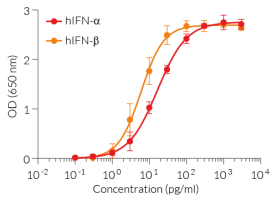
Recombinant human IFN-α2b is a high-quality and biologically active cytokine, validated using proprietary IFN-α/β reporter cells. This IFN-α subtype from the type I interferons family is produced in CHO cells to ensure protein glycosylation and bona fide 3D structure.
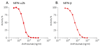
Recombinant human IFN-α2b can be used together with HEK-Blue™ IFN-α/β cells for the screening of inhibitory molecules, such as Rontalizumab and Aniftrolumab, two monoclonal antibodies targeting IFN-α and its receptor, respectively (see figures).
Key features
- Each lot is validated using HEK-Blue™ IFN-α/β cells
- Endotoxin < 1 EU/µg
- 0.22 µm sterile-filtered
Applications
- IFN-α2b detection and quantification assays (positive control)
- Screening and release assays for antibodies blocking IFN-α2b signaling
- Screening and release assays for engineered IFN-α2b proteins
Interferon alpha 2b (IFN-α2b) is the prototypic subtype of IFN-α used in fundamental research and most clinical applications. Despite its anti-viral protective effects, excessive IFN-α contributes to the pathogenesis of immune-mediated inflammatory diseases, including systemic lupus erythematosus (SLE) and STING-associated vasculopathy with onset in infancy (SAVI).
All InvivoGen products are for internal research use only, and not for human or veterinary use.
SPECIFICATIONS
Specifications
P01563
>108 IU/mg*
100 μg/ml in water
Phosphate buffer saline (pH 7.4), 5% saccharose, 2% HSA
0.22 µm filtration
The absence of bacterial contamination (e.g. lipoproteins and endotoxins) has been confirmed using HEK-Blue™ TLR2 and HEK‑Blue™ TLR4 cells.
Cellular assays (tested), ELISA
Each lot is functionally tested and validated.
*Please refer to the corresponding Certificate of Analysis (CoA) for the exact value.
CONTENTS
Contents
-
Product:Recombinant human IFN-α2b
-
Cat code:rcyc-hifna2b-1
-
Quantity:10 µg
1.5 ml endotoxin-free water
Shipping & Storage
- Shipping method: Room temperature
- -20°C
- Avoid repeated freeze-thaw cycles
Storage:
Caution:
Details
The human interferon-alpha family
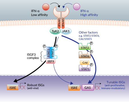
Type I interferons (IFN) include the IFN-α family, IFN-β, IFN-ε, IFN-κ, and IFN-ω. IFN-αs are important anti-viral cytokines that also have anti-proliferative and immuno-modulatory functions. The human IFN-α family comprises 13 genes encoding 12 proteins, with IFN-α13 being identical to IFN-α1. All IFN-αs bind to a common heterodimer receptor IFNAR1/IFNAR2. The ternary complex signals through the Janus kinase (JAK) and signal transducer and activator of the transcription (STAT) signaling pathway, inducing the formation of the ISGF3 transcriptional complex (STAT1/STAT2/IRF9). ISGF3 binds to IFN-stimulated response elements (ISRE) in the promoter regions of numerous IFN-stimulated genes (ISGs) [1].
Human IFN-α genes have evolved under strong selective pressure, suggesting a non-redundant role between IFN-α subtypes [2]. Although most studies have focused on IFN-α2 and IFN-α8, a consensus model for all IFN-αs has emerged depending on the affinity of a particular IFN-α subtype for IFNAR. Low-affinity IFN-α subtypes signal strictly through ISGF3 and induce robust ISGs, such as PKR, ISG56, and IFI16, which display anti-viral functions. Conversely, high-affinity IFN-α subtypes signal through ISGF3 and other factors, which activate “tunable” ISGs” such as CXCL10, IL-8, and ISG15, that induce anti-proliferative and immuno-modulatory functions [3]. IFN-α8, IFN-α10, and IFN-α14 have been identified as the most potent inducers of ISGs, while IFN-α1 is the weakest [4].
Human interferon-alpha 2b
The human interferon α2 (hIFN-α2) was the first highly active IFN subtype to be cloned and available for research. For this reason, hIFN-α2 has been the prototypic IFN-α among all other subtypes of this family used in fundamental research and most clinical applications [5, 6]. Human IFN-α2a and-α2b are allelic variants differing by a neutral lysine to arginine substitution at position 23 of the mature protein, respectively [5,6]. They are the only IFN-α subtypes with an O-glycosylation site (on Thr106) [6].
Human interferon-alpha 10
Natural human IFN-α10 is induced in peripheral blood mononuclear cells upon incubation with CpG-oligonucleotides and in plasmacytoid dendritic cells upon incubation with CpG-oligonucleotides or imiquimod [7]. This subtype of IFN-α is classified among the top three strongest ISG inducers [4, 8] with high anti-viral activity in vitro against human metapneumovirus [9] and hepatitis C virus (HCV) [10]. Human IFN-α10 displays a strong capacity to induce IFIT1, CXCL10, CXCL11, ISG15, and CCL8 [8]. Human IFN-α10 expression is IFN-α receptor-dependent as it is induced by other IFN-α subtypes upon infection with low doses of Sendai virus in vitro [11].
IFN-α subtypes available upon request for a minimum quantity : Please contact us
| Subtype | Alternate name | UnitProd ID | Source |
| IFN-α1 | IFN-alpha D | P01562 | CHO |
| IFN-α4a | IFN-alpha M1 | P05014 | CHO |
| IFN-α5 | IFN-alpha G | P01569 | CHO |
| IFN-α6 | IFN-alpha K | P05013 | CHO |
| IFN-α7 | IFN-alpha J1 | P01567 | CHO |
| IFN-α8 | IFN-alpha B2 | P31881 | CHO |
| IFN-α14 | IFN-alpha H2 | P01570 | HEK293 |
| IFN-α16 | IFN-alpha WA | P05015 | CHO |
| IFN-α17 | IFN-alpha I | P01571 | CHO |
| IFN-α21 | IFN-alpha F | P01568 | CHO |
References
1. Schreiber G. 2017. The molecular basis for differential type I interferon signaling. J. Biol. Chem. 292:7285-94.
2. Manry J. et al., 2011. Evolutionary genetic dissection of human interferons. J. Exp. Med. 208:2747-59.
3. Levin D. et al., 2014. Multifaceted activities of type I interferon are revealed by a receptor antagonist. Sci. Signal. 7(327). ra50.
4. Kurunganti S. et al., 2014. Production and characterization of thirteen human type-I interferon-α subtypes. Protein Expr. Purif. 103: 75-83.
5. Paul F. et al., 2015. IFNA2: The prototypic human interferon. Gene.
6. Antonelli G. et al., 2015. Twenty-five years of type I interferon-based treatment: A critical analysis of its therapeutic use. Cytokine Growth Factor Rev. 26(2):121-31.
7. Hillyer P. et al., 2012. Expression profiles of human interferon-alpha and interferon-lambda subtypes are ligand- and cell-dependent. Immunol. Cell. Biol. 90(8):774.
8. Moll H.P. et al., 2011. The differential activity of interferon-α subtypes is consistent among distinct target genes and cell types. Cytokines. 53:52.
9. Scagnolari C. et al., 2011. In vitro sensitivity of human metapneumovirus to type I interferons. Viral Immunol. 24(2):159.
10. Koyama T. et al., 2006. Divergent activities of interferon-alpha subtypes against intracellular hepatitis C virus replication. Hepatol. Res. 34(1):41.
11. Zaritsky L.A. et al., 2015. Virus multiplicity of infection affects type I interferon subtype induction profiles and interferon-stimulated genes. J. Virol. 89:11534.
DOCUMENTS
Documents
Technical Data Sheet
Safety Data Sheet
Validation Data Sheet
Certificate of analysis
Need a CoA ?


