


Gardiquimod™
-
Cat.code:
tlrl-gdqs-1
- Documents
ABOUT
TLR7 Agonist - Imidazoquinoline compound
Gardiquimod™ is an imidazoquinoline compound developed and manufactured by InvivoGen. Gardiquimod™ is a specific agonist for human and mouse Toll-like receptor 7 (TLR7). TLR7 and TLR8 are endosomal pattern recognition receptors that play an important role in the antiviral immune response [1].

Mode of action
Using our HEK-Blue™ reporter cell lines expressing human or mouse TLR7 or TLR8, we established that Gardiquimod™ is a human and mouse TLR7-specific agonist. Similar to Imiquimod, Gardiquimod™ induces the activation of NF-κB in HEK293 cells expressing human or mouse TLR7 (see figure). It is more potent than Imiquimod. Gardiquimod™ is a TLR7-specific ligand that does not activate TLR8; however, at high concentrations (>10 µg/ml), it might activate human, but not mouse TLR8.
Moreover, Gardiquimod™ is able to activate hTLR7-dependent NF-κB and IRF pathways, as assessed using our monocytic THP1-Dual™ reporter cell lines expressing two reporter genes, for the NF-κB-inducible SEAP and IRF-inducible Lucia luciferase, as well as hTLR7 or hTLR8 (see figure).
Key features of Gardiquimod™
- Specific activator of human and mouse TLR7
- Does not activate TLR8
- Each lot of Gardiquimod™ is highly pure (≥95%) and functionally tested
References:
1. Georg P. & Sander L.E., 2019. Innate sensors that regulate vaccine responses. Curr. Op. Immunol. 59:31.
All products are for research use only, and not for human or veterinary use.

SPECIFICATIONS
Specifications
TLR7
Human, Mouse
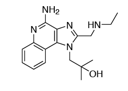
C17H23N5O • HCl
0.1 - 3 µg/ml
1 mg/ml in water
Negative (tested using EndotoxDetect™ assay)
Cellular assays
TLR7 activation
Each lot is functionally tested and validated.
CONTENTS
Contents
-
Product:Gardiquimod™
-
Cat code:tlrl-gdqs-1
-
Quantity:2 x 500 µg
1.5 ml of sterile endotoxin-free water
Shipping & Storage
- Shipping method: Room temperature
- -20°C
- Avoid repeated freeze-thaw cycles
Storage:
Caution:
Details
TLR7 and TLR8
TLR7 and TLR8 are endosomal pattern recognition receptors that share structural homology [1]. Both receptors are activated by single-stranded RNA (ssRNA) molecules, however, they exhibit different ligand-binding specificities and cellular expression patterns suggesting that they have nonredundant specialized roles.
TLR7 is essentially expressed by plasmacytoid dendritic cells (pDCs) but is also found in B cells and other myeloid cells [2] while TLR8 is highly expressed by myeloid cells and is absent from pDCs and B cells [2].
The endosomal distribution of TLR7 and TLR8 allows them to scan for the presence of microbial RNA in the phagocytic cargo. Their activation leads to NF-κB-, AP1-, and interferon regulatory factor (IRF)-mediated production of type I interferons (IFN-α/β) and pro-inflammatory cytokines [2].
Structural analyses have revealed that both TLR7 and TLR8 possess two binding sites (designated as Site 1 and Site 2) which do not share the same specificities.
Site 1 is highly conserved between TLR7 and TLR8 and binds nucleosides (guanosine (G) for TLR7 and uridine (U) for TLR8) or base analogs. The ligand preference for TLR7 and TLR8 is thus explained by the presence of specific residues in Site 1. Site 1 occupancy allows receptor dimerization and signaling.
Site 2 is less conserved and binds ssRNA with U(U) and U(G) motifs, respectively [3, 4]. Of note, ssRNA-binding to Site 2 is not sufficient for the formation of a signaling-competent TLR dimer but it strongly enhances the binding affinity of Site 1 [3, 4]. Thus, TLR7 and TLR8 appear to sense distinct RNA-degradation products rather than full-length ssRNAs [4].
1. Chuang T.H. & Ulevitch R.J., 2000. Cloning and characterization of a sub-family of human toll-like receptors: hTLR7, hTLR8, and hTLR9. Eur Cytokine Netw, 11:372-8.
2. Georg P. & Sander L.E., 2019. Innate sensors that regulate vaccine responses. Curr. Op. Immunol. 59:31.
3. Zhang Z. et al., 2018. Structural analyses of Toll-like receptor 7 reveal detailed RNA sequence specificity and recognition mechanism of agonistic ligands. Cell Rep. 25:3371.
4. Tanji H. et al., 2015. Toll-like receptor 8 senses degradation products of single-stranded RNA. Nat. Struct. Mol. Biol. 22:109.
Chemical structure of Gardiquimod™
DOCUMENTS
Documents
Technical Data Sheet
Validation Data Sheet
Safety Data Sheet
Certificate of analysis
Need a CoA ?


