


HeLa-Difluo™ hLC3 Cells
| Product | Unit size | Cat. code | Docs. | Qty. | Price | |
|---|---|---|---|---|---|---|
|
HeLa-Difluo™ hLC3 Cells Autophagy Reporter Cells |
Show product |
3-7 x 10e6 cells |
heldf-hlc3
|
Discontinued |
Notification: Reference #heldf-hlc3 has been discontinued - You might be interested in THP1-Difluo™ hLC3 Cells
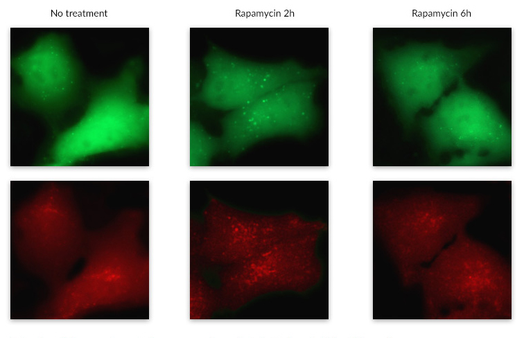
Monitoring autophagy activity
HeLa-Difluo™ hLC3 reporter cells are designed to monitor the autophagic flux by means of a fluorescent-labeled LC3B protein.
These cells, derived from the HeLa human epithelial carcinoma cell line, express the RFP::GFP::LC3 fusion protein where the human LC3B (microtubule-associated protein 1 light chain 3 beta) is fused to two fluorescent reporter proteins: RFP (acid-stable) and GFP (acid-sensitive). The LC3B protein is a good marker to assess the autophagic flux since it is recruited from the cytosol to be associated to all the autophagic structures: the nascent autophagosome (called the isolation membrane), the complete autophagosome and the autolysosome. Upon autophagy induction, diffuse cytoplasmic fluorescence shifts to autophagosomes and autolysosomes as fluorescent puncta.
Monitoring of autophagy activity relies on microscopy visualization of fluorescent puncta, progressive degradation of the GFP, and concurrent increase of RFP signal in autolysosomes. The number of fluorescent punctate structures per cell, the percentages of RFP-GFP positive and RFP positive cells can be used to assess autophagic flux as described previously [1,2].
HeLa-Difluo™ hLC3 cells are resistant to Zeocin®.
References
1. Loos B. et al. 2014. Defining and measuring autophagosome flux- concept and reality. Autophagy. 2014;10(11):2087-96.
2. Kimura s. et al., 2007. Dissection of the autophagosome maturation process by a novel reporter protein, tandem fluorescenttagged LC3. Autophagy. 3(5):452-60.
Figures

Specifications
Antibiotic resistance: Zeocin®
Growth medium: DMEM, 4.5 g/l glucose, 2 mM L-glutamine, 10% (v/v) fetal bovine serum (FBS), 100 U/ml penicillin, 100 μg/ml streptomycin, 100 μg/ml Normocin™.
Quality control:
- HeLa-Difluo™ hLC3 cells have been tested for their ability to respond to autophagic inducers.
- The stability of this cell line for 20 passages following thawing has been verified.
- HeLa-Difluo™ hLC3 cells are guaranteed mycoplasma-free.
Contents
- 1 vial of HeLa-Difluo™ hLC3 Cells (3-7 x 10⁶ cells)
- 100 μl Zeocin® (100 mg/ml).
- 1 ml Normocin™ (50 mg/ml). Normocin™ is a formulation of three antibiotics active against mycoplasmas, bacteria and fungi.
![]() Shipped on dry ice (Europe, USA, Canada and some areas in Asia)
Shipped on dry ice (Europe, USA, Canada and some areas in Asia)
See Data Sheet for storage conditions of each component.
Back to the top