


Recombinant anti-mouse CD4 antibody
| Product | Unit size | Cat. code | Docs. | Qty. | Price | |
|---|---|---|---|---|---|---|
|
Anti-mCD4-mIgG2a InvivoFit™ GK1.5-derived mouse monoclonal antibody against murine CD4 |
Show product |
1 mg 10 mg |
mcd4-mab10-1
|
|
Recombinant murinized CD4 antibody for in vivo use

InvivoGen’s engineered Anti-mCD4-mIgG2a InvivoFit™ antibody
Anti-mCD4-mIgG2a InvivoFit™ is a murinized anti-mouse monoclonal antibody (mAb) featuring the variable region of the previously described anti-mCD4 GK1.5 clone [1]. Using recombinant technology, the original GK1.5 rat IgG2b constant region has been replaced with a murine IgG2a format which mediates potent cytotoxic functions [2].
CD4 is a transmembrane protein primarily expressed on most thymocytes, and highly expressed by the peripheral mature CD4+ T cell population, including T helper (Th) and regulatory T (Treg) cells [3]. Other immune cells, such as monocytes and macrophages also express CD4, albeit to 10- to 20-fold fewer levels compared to T cells [4].
The anti-mCD4 GK1.5 mAb is commonly used for in vivo depletion of the CD4+ T cell population to study the role of this T cell subset in various immune responses, including anti-pathogenic responses, auto-immunity, cancer, or transplantation [5]. Depending on the nature of the experiment, extended treatment schedules (up to several months) may be required. Upon repeated injection of a xenogeneic mAb, mice produce anti–species antibodies, causing the removal of the administered mAb from circulation, thereby considerably reducing treatment efficacy. Moreover, this immunogenicity can lead to fatal hypersensitivity reactions [5-7] which can be reduced by mAb murinization [8].
Anti-mCD4-mIgG2a is provided in an InvivoFit™ grade, a high-quality standard specifically adapted to in vivo studies.
Key features of Anti-mCD4-mIgG2a InvivoFit™:
- Derives from the GK1.5 clone, rat IgG2b, κ
- Features the mIgG2a isotype (constant region)
- Filter-sterilized (0.2 µm), endotoxin level < 1 EU/mg
- Suitable for parental delivery in mice (azide-free)
- Low aggregation < 5%
- Produced in animal-free facilities and defined media
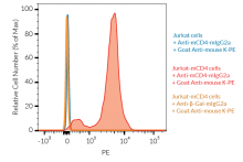
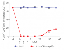
Anti-mCD4-mIgG2a InvivoFit™ is produced in Chinese hamster ovary (CHO) cells and purified by affinity chromatography with protein A. The specific binding of this mAb to cell surface mCD4 and its in vivo depleting function have been confirmed (see Figures).
References:
1. Dialynas D.P. et al., 1983. Characterization of the murine antigenic determinant, designated L3T4a, recognized by monoclonal antibody GK1.5: expression of L3T4a by functional T cell clones appears to correlate primarily with class II MHC antigen-reactivity. Immunol Rev. 74:29-56
2. Nimmerjahn F. & Ravetch J.V., 2005. Divergent immunoglobulin g subclass activity through selective Fc receptor binding. Science. 310(5753):1510-2.
3. Zhu J. et al., 2010. Differentiation of effector CD4 T cell populations. Annual Rev Immunol. 28:445-489.
4. Collman R. et al., 1990. Macrophage-tropic strains of human immunodeficiency virus type 1 utilize the CD4 receptor. J Virol. 64(9):4468-76.
5. Laky K. & Kruisbeek A.M., 2016. In vivo depletion of T lymphocytes. Current Protocols Immunology. 4.1.1-4.1.9. doi: 10.1002/0471142735.im0401s113.
6. Mall C. et al., 2016. Repeated PD-1/PD-L1 monoclonal antibody administration induces fatal xenogenic hypersensitivity reactions in a murine model of breast cancer. Onco Immunol. 5(2):e1075114.
7. Murphy, J.T. et al., 2014. Anaphylaxis caused by repetitive doses of a GITR agonist monoclonal antibody in mice. Blood. 123(14):2172-2180.
8. Belmar N.A. et al., 2017. Murinization and H chain isotype matching of Anti-GITR antibody DTA-1 reduces immunogenicity-mediated anaphylaxis in C57BL/6 mice. J Immunol. 198:4502-4512.
Figures

Specifications
Specificity: Targets cells expressing murine CD4
Formulation: Lyophilized from 0.2 μm filtered solution in 150 mM sodium chloride, 20 mM sodium phosphate buffer with 5% saccharose
Clonality: Monoclonal antibody
Isotype: Murine IgG2a, kappa
Control: mIgG2a InvivoFit™ isotype control
Source: CHO cells
Purity: Purified by affinity chromatography with protein A
Tested application: Flow cytometry; in vivo depletion
Quality control:
- The binding of Anti-mCD4-mIgG2a InvivoFit™ to mCD4 has been confirmed using Flow cytometry
- Mouse CD4+ T cell in vivo depletion using Anti-mCD4-mIgG2a InvivoFit™ has been confirmed
- The complete sequence of the antibody construct has been verified
- < 5% aggregates (confirmed by size exclusion chromatography)
- Endotoxin level <1 EU/mg (determined by the LAL assay)
Contents
Anti-mCD4-mIgG2a InvivoFit™ is filter-sterilized (0.2 µm), endotoxin-free, azide-free, and lyophilized.
This product is available in two pack sizes:
- mcd4-mab10-1: 1 mg
- mcd4-mab10-10: 10 mg
![]() The product is shipped at room temperature.
The product is shipped at room temperature.
![]() Store lyophilized antibody at -20 °C.
Store lyophilized antibody at -20 °C.
![]() Lyophilized product is stable for at least 1 year
Lyophilized product is stable for at least 1 year
![]() Avoid repeated freeze-thaw cycles.
Avoid repeated freeze-thaw cycles.
InvivoFit™
InvivoFit™ is a high-quality standard specifically adapted for in vivo studies. InvivoFit™ products are filter-sterilized (0.2 µm) and filled under strict aseptic conditions in a clean room. The level of bacterial contaminants (endotoxins and lipoproteins) in each lot is verified using a LAL assay and a TLR2 and TLR4 reporter assay.
Back to the topDetails
CD4 Background:
The cluster of differentiation 4 (CD4) receptor (formerly named L3T4, or T4) is a 55 kDa transmembrane protein primarily expressed on most thymocytes, and highly expressed by the peripheral mature CD4+ T cell population, including T helper (Th) and regulatory T (Treg) cells [1]. Other immune cells, such as monocytes and macrophages also express CD4, albeit to 10- to 20-fold fewer levels compared to T cells [2]. Besides its role in the positive selection and development of CD4+ T cells, the CD4 receptor plays a critical role during their activation. It fulfills an intercellular adhesion function by interacting with the α2 or β2 domain of MHC class II molecules, thereby stabilizing the interaction between the TCR on the T cell and the MHC-peptide complex on the antigen-presenting cell [3]. Upon antigen recognition, the proximity association of CD4 and the TCR/CD3 complex on T cells triggers downstream intracellular signaling and participates in the T helper differentiation [1].
References:
1. Zhu J. et al., 2010. Differentiation of effector CD4 T cell populations. Annual Rev Immunol. 28:445-489.
2. Collman R. et al., 1990. Macrophage-tropic strains of human immunodeficiency virus type 1 utilize the CD4 receptor. J Virol. 64(9):4468-76.
3. Doyle C. & Strominger J.L., 1987. Interaction between CD4 and class II MHC molecules mediates cell adhesion. Nature 330:256-259.