


Recombinant anti-mouse IL-1β antibody
| Product | Unit size | Cat. code | Docs. | Qty. | Price | |
|---|---|---|---|---|---|---|
|
Anti-mIL-1β-mIgG1 Recombinant mouse mAb against mIL-1β (clone 7E3). For in vitro use. |
Show product |
200 µg |
mil1b-mab9-02
|
|
||
|
Anti-mIL-1β-mIgG1 InvivoFit™ Recombinant mouse mAb against mIL-1β (clone 7E3). For in vivo use. |
Show product |
1 mg 10 mg |
mil1b-mab9-1
|
|
Recombinant mouse IL-1 beta antibody

Neutralizing monoclonal antibody against murine IL-1β
InvivoGen provides a recombinant fully mouse anti-mouse IL-1β monoclonal antibody (mAb) that was previously extracted from hybridoma. It is now expressed and produced in Chinese hamster ovary (CHO) cells, ensuring reliability and lot-to-lot reproducibility. Thereby, common hybridoma-related drawbacks such as the generation of non-relevant mAbs containing aberrant light chains are avoided [1]. The sequence of the Anti-mIL-1β-mIgG1 is 100% murine (constant and variable regions), as the original clone (clone 7E3) was raised in mice using a proprietary method. This feature allows for reduced immunogenicity and risks of fatal hypersensitivity reactions upon repeated mAb injections into mice [2-4].
InvivoGen provides this antibody in two grades:
- In vitro use: Anti-mIL-1β-mIgG1
- In vivo use: Anti-mIL-1β-mIgG1 InvivoFit™
All InvivoFit™ products are handled in a clean room, filter-sterilized, and tested for bacterial contaminants. Additionally, this grade guarantees a low levels of endotoxins (<1 EU/ml). The buffer formulation is specifically adapted for in vivo studies.
Key features:
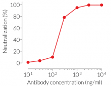
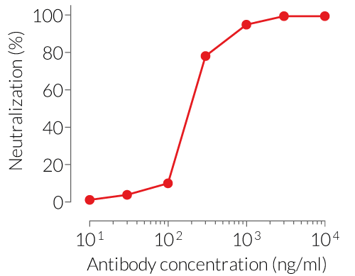
- Potent and specific neutralizing activity against mIL-1β (see figure)
- Sequence is 100% murine
- Murine IgG1 isotype (constant region)
- Free from non-relevant mAbs found in hybridoma-based productions
- Produced in animal-free facilities and defined media
- Low aggregation <5%
-
InvivoFit™ grade is available
Anti-mIL-1β-mIgG1 is designed to efficiently neutralize the biological activity of mIL-1β. Interleukin 1β (IL-1β) is a soluble pro-inflammatory cytokine that plays a critical role in the host’s response to infection and injury [5].
References:
1. Bradbury, A. et al. 2018. When monoclonal antibodies are not monospecific: Hybridomas frequently express additional functional variable regions. mAbs, 10(4), 539–546
2. Mall C. et al., 2016. Repeated PD-1/PD-L1 monoclonal antibody administration induces fatal xenogenic hypersensitivity reactions in a murine model of breast cancer. Onco Immunol. 5(2):e1075114.
3. Murphy, J.T. et al., 2014. Anaphylaxis caused by repetitive doses of a GITR agonist monoclonal antibody in mice. Blood. 123(14):2172-2180.
4. Belmar N.A. et al., 2017. Murinization and H chain isotype matching of Anti-GITR antibody DTA-1 reduces immunogenicity-mediated anaphylaxis in C57BL/6 mice. J Immunol. 198:4502-4512.
5. Dinarello CA., 2011. Interleukin-1 in the pathogenesis and treatment of inflammatory diseases. Blood. 117:3720–3732.
Figures
Specifications
Target: Murine IL-1β (mIL-1β)
Specificity: No cross-reactivity with human IL-1β
Clone: 7E3
Source: CHO cells
Isotype: Murine IgG1, kappa
Control: Murine IgG1 Isotype Control
Formulation of Anti-mIL-1β-mIgG1: Lyophilized from 0.2 µm filtered solution in a sodium phosphate buffer with glycine, saccharose, and stabilizing agents
Formulation of Anti-mIL-1β-mIgG1 InvivoFit™: Lyophilized from 0.2 µm filtered solution in a sodium phosphate buffer with 5% saccharose
Tested applications Blocking & Neutralization
Quality control:
- These products have been validated using neutralization cellular assays.
- The complete sequence of these antibodies has been verified.
- The absence of bacterial contamination (e.g. lipoproteins and endotoxins) has been confirmed using HEK-Blue™ TLR2 and HEK‑Blue™ TLR4 cells.
- The endotoxin level in Anti-mIL-1β-mIgG1 InvivoFit™ is <1 EU/mg (determined by a LAL assay).
Contents
Please note: each mAb is sold separately.
Anti-mIL-1β-mIgG1
- mil1b-mab9-02: 200 µg, lyophilized
Anti-mIL-1β-mIgG1 InvivoFit™
- mil1b-mab9-1: 1 mg, lyophilized
-
mil1b-mab9-10: 10 mg, lyophilized
![]() The product is shipped at room temperature.
The product is shipped at room temperature.
![]() Store lyophilized antibody at -20 °C.
Store lyophilized antibody at -20 °C.
![]() Lyophilized product is stable for at least 1 year.
Lyophilized product is stable for at least 1 year.
![]() Avoid repeated freeze-thaw cycles.
Avoid repeated freeze-thaw cycles.
InvivoFit™
InvivoFit™ is a high-quality standard specifically adapted for in vivo studies. InvivoFit™ products are filter-sterilized (0.2 µm) and filled under strict aseptic conditions in a clean room. The level of bacterial contaminants (endotoxins and lipoproteins) in each lot is verified using a LAL assay and a TLR2 and TLR4 reporter assay.
Back to the topDetails
Interleukin-1 beta (IL-1β) is a potent pro-inflammatory cytokine involved in a variety of cellular activities including cell migration, T lymphocyte activation and pro-inflammatory cytokine production [1]. Its inactive precursor, pro-IL-1β, is secreted by immune cells, notably the innate system as well as epithelia cells in response to stimuli such as inflammation, infection or cell damage. In order to get activated, pro-IL-1β is cleaved by caspase-1 upon inflammasome activation [1,2].
Following caspase-1-dependent processing, the mature IL-1β is rapidly secreted through pores of the plasma membrane. There, it interacts with the IL-1 receptor 1 (IL-1R1), which is shared with IL-1α. Upon receptor-ligand binding, a structural change of the receptor occurs allowing the co-receptor IL-1R3 to bind and form a trimeric complex. Subsequently, the intracellular adapter protein MyD88 (myeloid differentiation primary response 88) becomes phosphorylated triggering an intricate sequence of (auto-) phosphorylation, complex formation and ubiquitination events. Finally, activated NK-kB, c-Jun N-terminal kinase (JNK) and p38 mitogen-activated protein kinase (MAPK) pathways induce the expression of inflammatory cytokines and chemokines, such as IL-6 and IL-8 [3]. Due to its role in mediating acute and chronic inflammation, IL-1β has emerged as a therapeutic target for auto-inflammatory diseases [4].
References:
1. O’Neill L., 2008. The interleukin-1 receptor/Toll-like receptor superfamily: 10 years of progress. Immunol. Rev. 226, 10–18. 4. 2.
2. Dinarello C., 2018. Overview of the IL-1 family in innate inflammation and acquired immunity. Immunol Rev. 281(1): 8–27.
3. Weber A. et al., 2010. Interleukin-1 (IL-1) pathway. Sci Signal. 3(105).
4. Dinarello CA., 2011. Interleukin-1 in the pathogenesis and treatment of inflammatory diseases. Blood. 117:3720–3732.